The 3D models on this website are completely interactive! If you're using a mobile device, you can view them in augmented reality.
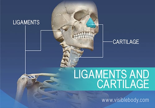
What is the skeletal system made of? What does the skeletal system do? At the simplest level, the skeleton is the framework that provides structure to the rest of the body and facilitates movement. The skeletal system includes over 200 bones, cartilage, and ligaments.
The human skeleton has a number of functions, such as protection and supporting weight. Different types of bones have differing shapes related to their particular function.
So, what are the different types of bones? How are they categorized?
There are five types of bones in the skeleton: flat, long, short, irregular, and sesamoid.
Let’s go through each type and see examples.
The bones of the human skeleton are divided into two groups. The appendicular skeleton includes all the bones that form the upper and lower limbs, and the shoulder and pelvic girdles. The axial skeleton includes all the bones along the body’s long axis. Let’s work our way down this axis to learn about these structures and the bones that form them.
The bones of the human skeleton are divided into two groups. The axial skeleton includes all the bones (that form bony structures) along the body’s long axis. The bones of the appendicular skeleton make up the rest of the skeleton, and are so called because they are appendages of the axial skeleton. The appendicular skeleton includes the bones of the shoulder girdle, the upper limbs, the pelvic girdle, and the lower limbs.
Joints hold the skeleton together and support movement. There are two ways to categorize joints. The first is by joint function,also referred to as range of motion. The second way to categorize joints is by the material that holds the bones of the joints together; that is an organization of joints by structure.
Joints in the human skeleton can be grouped by function (range of motion) and by structure (material). Here are some joints and their categorizations.
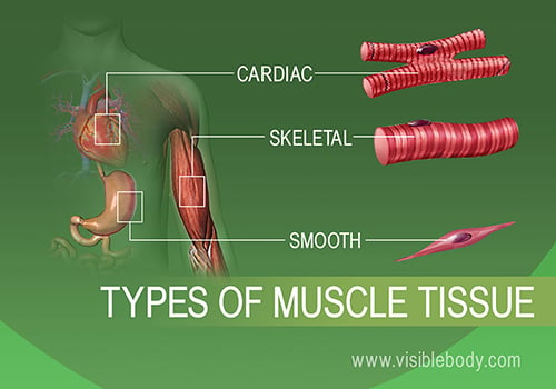
At the simplest level, muscles allow us to move. Smooth muscle and cardiac muscle move to facilitate body functions like heartbeats and digestion. The movement of these muscles is directed by the autonomic part of the nervous system—those are the nerves that control organs. Skeletal muscles move our bodies in space. They take direct instruction from the specific nerves that innervate each muscle. Want to learn more about the muscles in the human body? Here are five other facts to keep in mind about the muscular system.
About half of your body’s weight is muscle. In the muscular system, muscle tissue is categorized into three distinct types: skeletal, cardiac, and smooth. Each type of muscle tissue in the human body has a unique structure and a specific role. Skeletal muscle moves bones and other structures. Cardiac muscle contracts the heart to pump blood. The smooth muscle tissue that forms organs like the stomach and bladder changes shape to facilitate bodily functions. Here are more details about the structure and function of each type of muscle tissue in the human muscular system.
There are over 600 muscles in the human body. Learning the muscular system often involves memorizing details about each muscle, like where a muscle attaches to bones and how a muscle helps move a joint. In textbooks and lectures these details about muscles are described using specialized vocabulary that is hard to understand. Here is an example: The triceps brachii has three bellies with varying origins (scapula and humerus) and one insertion (ulna). It is a prime mover of elbow extension. The anconeus acts as a synergist in elbow extension.
How do the bones of the human skeleton move? Skeletal muscles contract and relax to mechanically move the body. Messages from the nervous system cause these muscle contractions. The whole process is called the mechanism of muscle contraction and it can be summarized in three steps:
Muscles allow us to move, but sometimes the wear and tear that comes from moving our bodies can lead to disorders of the muscular system. Below are some of the most common muscular pathologies.
The carpal tunnel is the passageway in the wrist where the median nerve and flexor tendons pass through a narrow opening. Carpal tunnel syndrome, which is also called median nerve compression, occurs when the tendons become inflamed, causing compression of the median nerve. Symptoms include pain, numbness, and eventual weakness in the hand. Carpal tunnel syndrome can occur for a variety of reasons including hereditary predisposition, repetitive movements, diabetes, or thyroid disorders.
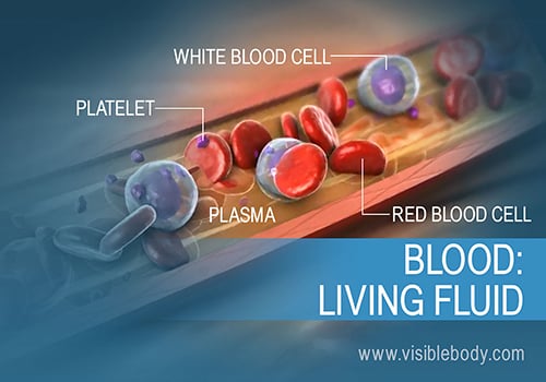
The heart pumps blood through a vast network of arteries and veins. Blood is a living fluid. It transports oxygen and other essential substances throughout the body, fights sickness, and performs other vital functions. Below are 8 important facts about blood.
The heart is a hollow, muscular organ that pumps oxygenated blood throughout the body and deoxygenated blood to the lungs. This key circulatory system structure is comprised of four chambers. One chamber on the right receives blood with waste (from the body) and another chamber pumps it out toward the lungs where the waste is exhaled. One chamber on the left receives oxygen-rich blood from the lungs and another pumps that nutrient-rich blood into the body. Two valves control blood flow within the heart’s chambers, and two valves control blood flow out of the heart.
Blood flowing through the circulatory system transports nutrients, oxygen, and water to cells throughout the body. The journey might begin and end with the heart, but the blood vessels reach every vital spot along the way. These arteries, veins, and capillaries make for a vast network of pipes. If you were to lay out all the blood vessels of the body in a line, they would stretch for nearly 60,000 miles. That’s enough to circle the earth almost three times!
Blood must always circulate to sustain life. It carries oxygen from the air we breathe to cells throughout the body. The pumping of the heart drives this blood flow through the arteries, capillaries, and veins. One set of blood vessels circulates blood through the lungs for gas exchange. The other vessels fuel the rest of the body. Read on to learn more about these crucial circulatory system functions.
The circulatory system, also called the cardiovascular system, includes the heart and the network of blood vessels that circulate blood throughout the body. Several diseases and disorders can affect this system. Below are some of the most common pathologies.
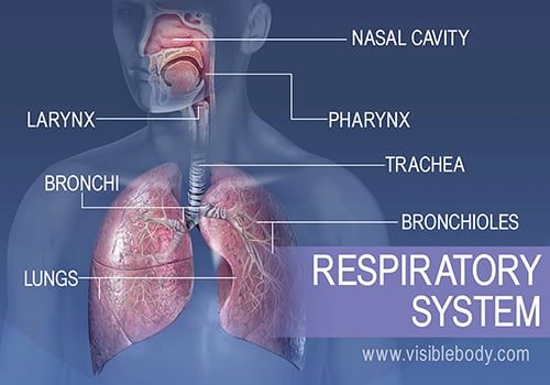
Through breathing, inhalation and exhalation, the respiratory system facilitates the exchange of gases between the air and the blood and between the blood and the body’s cells. The respiratory system also helps us to smell and create sound. The following are the five key functions of the respiratory system.
The upper respiratory system, or upper respiratory tract, consists of the nose and nasal cavity, the pharynx, and the larynx. These structures allow us to breathe and speak. They warm and clean the air we inhale: mucous membranes lining upper respiratory structures trap some foreign particles, including smoke and other pollutants, before the air travels down to the lungs.
The lower respiratory system, or lower respiratory tract, consists of the trachea, the bronchi and bronchioles, and the alveoli, which make up the lungs. These structures pull in air from the upper respiratory system, absorb the oxygen, and release carbon dioxide in exchange. Other structures, namely the thoracic cage (or rib cage) and the diaphragm, protect and support these functions.
There are two types of respiratory diseases and disorders: infectious and chronic. Pulmonary infections are most commonly bacterial or viral. In the viral type, a pathogen replicates inside a cell and causes a disease, such as the flu. Chronic diseases, such as asthma, are persistent and long-lasting. They can relapse and the patient can go into remission, only to suffer symptoms again at a later time.
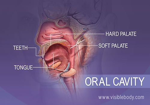
The digestive system is a kind of processing plant inside the body. It takes in food and pushes it through organs and structures where the processing happens. The fuels and nutrients we need are extracted, and the digestive system discards the rest.
The oral cavity is bounded by the teeth, tongue, hard palate, and soft palate. These structures make up the mouth and play a key role in the first step of digestion: ingestion. This is where the teeth and tongue work with salivary glands to break down food into small masses that can be swallowed, preparing them for the journey through the alimentary canal.
The alimentary canal is a single continuous tube that includes the oral cavity, pharynx, esophagus, stomach, small intestine, and large intestine. After food is chewed, made into a bolus, and swallowed, the action of the epiglottis routes the bolus into the esophagus. From there, peristaltic waves propel ingested foodstuffs through the alimentary canal.
Food that is chewed in the oral cavity then swallowed ends up in the stomach where it is further digested so its nutrients can be absorbed in the small intestine. The salivary glands, liver and gall bladder, and the pancreas aid the processes of ingestion, digestion, and absorption. These accessory organs of digestion play key roles in the digestive process. Each of these organs either secretes or stores substances that pass through ducts into the alimentary canal.
Ingested food is chewed, swallowed, and passes through the esophagus into the stomach where it is broken down into a liquid called chyme. Chyme passes from the stomach into the duodenum. There it mixes with bile and pancreatic juices that further break down nutrients. Finger-like projections called villi line the interior wall of the small intestine and absorb most of the nutrients. The remaining chyme and water pass to the large intestine, which completes absorption and eliminates waste.
Diseases and disorders of the digestive system can involve infection or damage to organs and other tissues and structures. They may also affect actions of the digestive system, such as the sealing of the esophagus from stomach acids or the free flow of fluids through the bile ducts. Symptoms can arise during digestion or may be chronic.
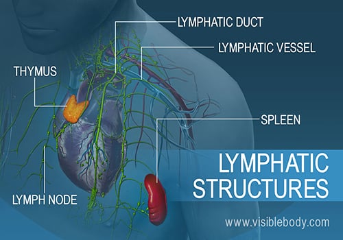
The lymphatic system includes a network of vessels, ducts, and nodes, as well as organs and diffuse tissue that support the circulatory system. These structures help to filter harmful substances from the bloodstream. Organs of the lymphatic system, such as the spleen, thymus, and tonsils, house specialized cells that destroy the harmful pathogens.
Immunity is the body’s defense system against infection and disease. White blood cells play a key role. Some rush to attack any harmful microbes that invade the body. Other white blood cells become specialists, adapted to fight particular pathogens. All of them work to keep the body as healthy as possible.
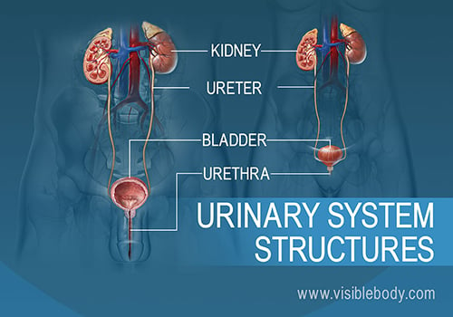
The kidneys, ureters, bladder, and urethra are the primary structures of the urinary system. They filter blood and remove waste from the body in the form of urine. The size and position of lower urinary structures vary with male and female anatomy.
The kidneys sit at the back of the abdominal wall and at the start of the urinary system. These organs are constantly at work: Nephrons, tiny structures in the renal pyramids, filter gallons of blood each day. The kidneys reabsorb vital substances, remove unwanted ones, and return the filtered blood back to the body. As if they weren’t busy enough, the kidneys also create urine to remove all the waste.
The kidneys filter unwanted substances from the blood and produce urine to excrete them. There are three main steps of urine formation: glomerular filtration, reabsorption, and secretion. These processes ensure that only waste and excess water are removed from the body.
Urine produced in the kidneys travels down the ureters into the urinary bladder. The bladder expands like an elastic sac to hold more urine. As it reaches capacity, the process of micturition, or urination, begins. Involuntary muscle movements send signals to the nervous system, putting the decision to urinate under conscious control.
Diseases of the kidneys or bladder can compromise urinary system functions. Below are some common diseases of the urinary system.
The kidneys produce urine to eliminate waste. Kidney stones can form when mineral and acid salts in the urine crystallize and stick together. If the stone is small, it can pass easily through the urinary system and out of the body. A larger stone can get stuck in the urinary tract, however. A stuck kidney stone causes pain and can block the flow of urine.
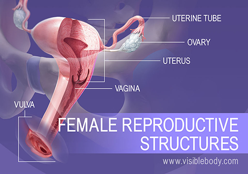
The female reproductive system includes external and internal genitalia. The vulva and its structures form the external genitalia. The internal genitalia include a three-part system of ducts: the uterine tubes, the uterus, and the vagina. This system of ducts connects to the ovaries, the primary reproductive organs. The ovaries produce egg cells and release them for fertilization. Fertilized eggs develop inside the uterus.
The testes are the primary reproductive organs and generate sperm cells through a process called spermatogenesis. The glands of the male reproductive system produce sperm and seminal fluid. The prostate gland, the seminal vesicles, and the bulbourethral glands contribute seminal fluid to semen, which carries and protects the sperm. During sexual intercourse, semen moves through a series of ducts to deliver the semen directly into the female reproductive system.
In the reproductive process, a male sperm and a female egg provide the information required to produce another human being. Conception occurs when these cells join as the egg is fertilized. Pregnancy begins once the fertilized egg implants in the uterus. The embryo grows and becomes surrounded by structures that provide support and nourishment. Eyes, limbs, and organs appear as the embryo develops into a fetus. The fetus grows inside the uterus until pregnancy ends with labor and birth. By then all body systems are in place—including the reproductive system that can one day help produce another human being.
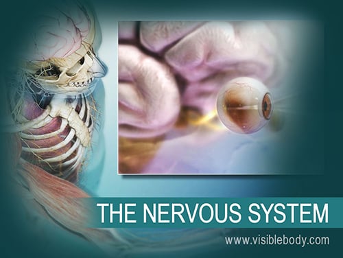
The nervous system is the most complex and highly organized body system. It receives information from the sensory organs via nerves, transmits the information through the spinal cord, and processes it in the brain. The nervous system directs our body’s reactions to the world and also controls most of our internal functions, everything from muscle movement and blood vessel dilation to the learning of anatomy and physiology facts. How does it manage all this? By sending lightning-quick signals, electrical and chemical, between cells.
The most complex workings of the nervous system depend on messages sent through neurons. Together with their support cells, neuroglia, neurons make up all nervous system tissue. They receive and relay messages quickly, conducting them as electrical signals. Neurons release neurotransmitters, chemicals that jump the message to the next neuron or body cell. Specialized neurons in the spinal cord form neural tracts that speed messages to and from the brain. Read on to learn more about these “live-wire” cells.
The brain directs our body’s internal functions. It also integrates sensory impulses and information to form perceptions, thoughts, and memories. The brain gives us self-awareness and the ability to speak and move in the world. Its four major regions make this possible: The cerebrum, with its cerebral cortex, gives us conscious control of our actions. The diencephalon mediates sensations, manages emotions, and commands whole internal systems. The cerebellum adjusts body movements, speech coordination, and balance, while the brain stem relays signals from the spinal cord and directs basic internal functions and reflexes.
The nervous system must receive and process information about the world outside in order to react, communicate, and keep the body healthy and safe. Much of this information comes through the sensory organs: the eyes, ears, nose, tongue, and skin. Specialized cells and tissues within these organs receive raw stimuli and translate them into signals the nervous system can use. Nerves relay the signals to the brain, which interprets them as sight (vision), sound (hearing), smell (olfaction), taste (gustation), and touch (tactile perception).
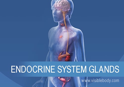
The glands of the endocrine system secrete hormones into the bloodstream to maintain homeostasis and regulate metabolism. The hypothalamus and the pituitary gland are the command and control centers, directing hormones to other glands and throughout the body. Other primary endocrine glands, including the thyroid and parathyroid glands, the adrenal glands, and the pineal gland, adjust the levels of various substances in the blood and regulate metabolism, growth, the sleep cycle, and other processes. Organs such as the pancreas also secrete hormones as part of the endocrine system. Secondary endocrine organs include the gonads, kidneys, and thymus.
Hormones regulate internal functions from metabolism and growth to sexual development and the induction of birth. They circulate through the bloodstream, bind to target cells, and adjust the function of whole tissues and organs. It all starts with the hypothalamus and the pituitary gland, the masters of the endocrine system. The hormones they release control the secretions of the other endocrine glands and most endocrine functions. Throughout the body, hormones enable reactions to stress and other outside changes and keep regular processes running smoothly.
When you select "Subscribe" you will start receiving our email newsletter. Use the links at the bottom of any email to manage the type of emails you receive or to unsubscribe. See our privacy policy for additional details.