Why Visible Body’s Models Are a Step Above the Competition
Posted on 10/25/24 by The Team at Visible Body
There are several anatomy and physiology visualization apps on the market today, so when instructors and administrators first encounter Visible Body Suite or Courseware, they often ask the same question: What makes Visible Body’s models so special?
In this blog post, we’ll provide the answer with five reasons why Visible Body’s models stand out over our competition.
1. Our anatomy models are created by experts
All of Visible Body’s 3D models are created by medical artists with degrees in biomedical illustration.
As far as we are aware, Visible Body is the only company that employs medical artists academically trained in biomedical illustration.
Having specialists means we can produce a more accurate product. For example, let’s look at our trachea model, paying attention to where it bifurcates into the left and right primary bronchus.
In Visible Body’s model, the left primary bronchus takes a hard turn and lies almost horizontally.
Why does the angle matter? For one, the vertical nature of the right primary bronchus has clinical significance. If a child aspirates an object and is rushed into the ER, the clinicians know that the foreign body will most likely end up in the right primary bronchus.

Example case study illustrating the clinical significance of the left primary bronchus. GIF from VB Suite.
Additionally, the Visible Body model shows the spatial relationships between the bronchi and the heart—the left primary bronchus is at such an angle because it rests below the aortic arch.

GIF from VB Suite.
We’ve noticed that some of our competitors’ trachea models ignore this nuance for a simpler, less accurate model. Instead, the trachea and primary bronchi appear like an inverted Y, with both primary bronchi jutting out at the same angle.
Spatial relationships between structures matter. Our team brings the expertise needed to create accurate models so that the next generation of medical professionals can better visualize the body and deliver quality patient care.
2. Our art style is both identifiable and realistic
Another way our medical artists’ expertise shines is through Visible Body’s model style.
When it comes to 3D anatomy models, artists must create a balance between realism and identifiable structures. In real life, the colors of various organs are often too similar to identify different structures. However, if an artist overcorrects on making structures identifiable, the models look plasticky and artificial.
Visible Body uses different textures and colors to make each structure identifiable without losing realism.

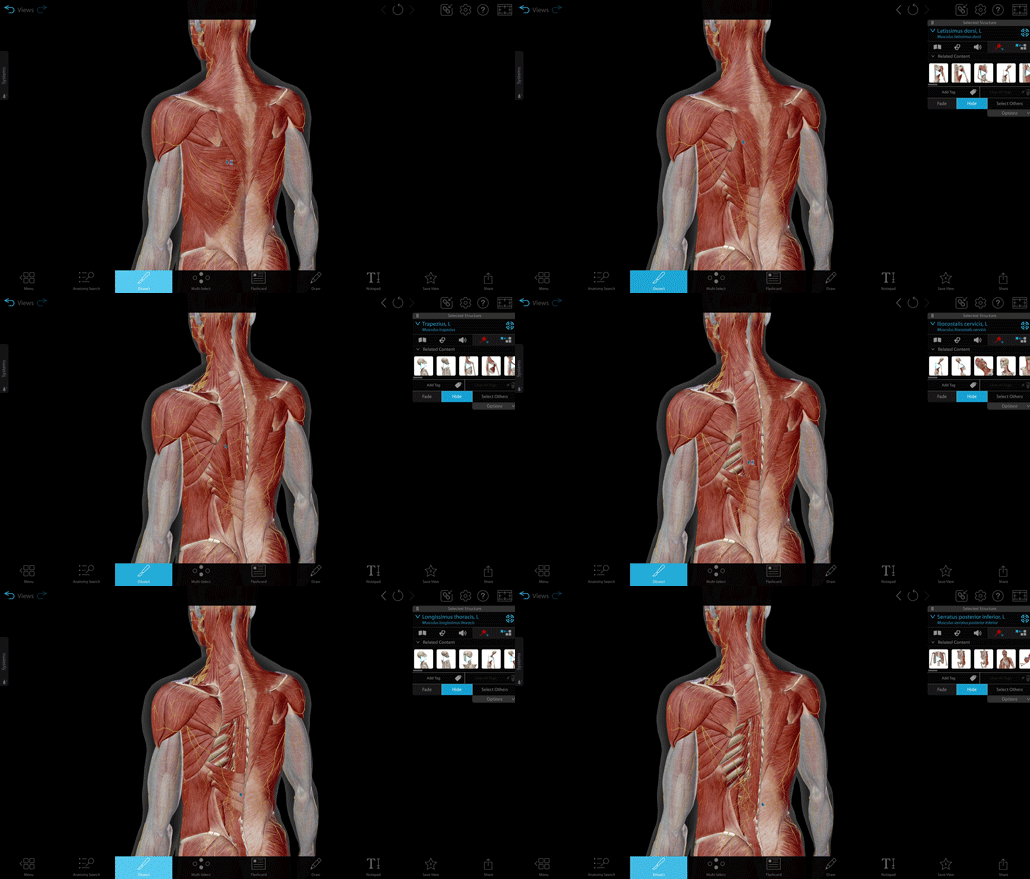
Simulated dissection of back muscles. GIF from VB Suite.
There are over 600 muscles in the human body, and striation looks different in every one of them. Instead of stamping every muscle with the same texture, each muscle in VB Suite has its own texture map.
It’s our goal that our models’ quality and details are such that you could use any screenshot as an illustration.
3. We focus on educational goals
Visible Body is focused on helping instructors teach and students learn, so we take pedagogy seriously. We’re not interested in making models just for the “cool factor”—our models align with learning goals and help students master content faster.
For example, let’s look at the pancreatic acini and islets microanatomy model. The educational goal? Students should identify the key cells in the pancreas.

Image from VB Suite.
This model uses exaggerated perspective and foreshortening to add focus on the cells of the pancreatic acinus and islet pancreatic model. The tail of the pancreas is present in the background, less emphasized but still present to add context.
As they navigate the model, students start with a “zoomed in” view that shows the structures inside the pancreatic acinar cells, the centroacinar cells, and the alpha, beta, delta, and PP cells within the pancreatic islet. Then, students view the shape of the pancreatic lobule and its relationship to the intercalated ducts and the pancreatic branch of the splenic artery.
In presenting the cells, this model also presents part of the pancreas, adding reference and orientation. Most of the islets are in the body and tail of the pancreas, so this inclusion also acts as a visual cue.
Visible Body’s artists leverage orientation, textures, colors, and spatial relationships to emphasize certain elements so that students get the most out of our anatomy models.
4. Our models match course topics
Visible Body follows A&P courses to ensure that our models align with how instructors actually teach.
We also correlate with popular A&P textbooks to make sure that when instructors switch from textbook to Visible Body, they won’t miss out on any of the visuals they love using in their courses. Instead, those visuals have been enhanced in 3D!
Visible Body works with instructor-reviewers to ensure that all content we create has value in the classroom.
For more on Visible Body’s content development process, check out this blog post.
5. Students can learn in multiple modalities
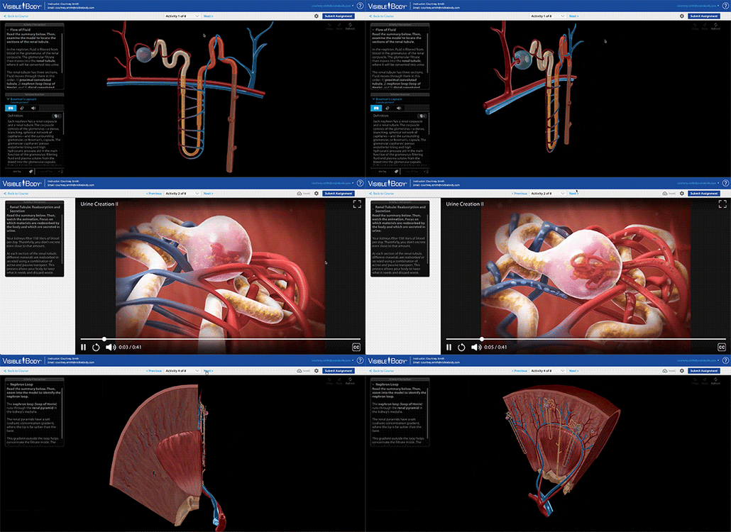
We’ve talked a lot so far about Visible Body’s amazing 3D gross and microanatomy models, but in VB Suite and Courseware, you’ll also find animations, illustrations, and histology slides.

GIF showing an assignment in Courseware.
When we create content, we think about the best way to visually express concepts. Sometimes, that results in a 3D model, and other times, we create an animation or illustration because information is conveyed better in those modalities. Like we've said before, we don’t focus on being flashy—we focus on teaching.
Incorporating multiple modalities is a tried-and-true pedagogical approach that engages students and challenges them to make connections across concepts.
Visible Body focuses on quality education
Our models reflect our trained medical artists’ expertise, medical accuracy, and an emphasis on pedagogy—that’s what sets us apart from the competition.
To see the difference for yourself, sign up for a free instructor trial!
You may also be interested in these blog posts:
- Why Do Librarians Love Visible Body Suite?
- A collection of comparisons between Visible Body Courseware and competitors
- Plastic Models + VB Suite: New Research Examines Engagement and Interest in A&P


Be sure to subscribe to the Visible Body Blog for more anatomy awesomeness!
Are you an instructor? We have award-winning 3D products and resources for your anatomy and physiology course! Learn more here.


