Using 3D Anatomy Models to Teach in the Lab and Online: An Interview with Prof. Jay Gump
Posted on 4/24/20 by Laura Snider
Prof. Jay Gump, a senior lecturer in the Kinesiology program at the University of Massachusetts, has a few years experience using 3D models in lab, lecturing online, and assigning online anatomy lab homework.

Jay was kind enough to spend an hour with the VB team sharing his experiences using technology in the lab and moving anatomy learning online. We’ve collected some of the most important excerpts for you here. (Also, the text has been edited for clarity.)
Jay's background
Education: PhD in Physiology from University of Vermont, post-doc at UCSF.
How he got into teaching: A friend at Greenfield Community College asked him to teach a course.
"I loved it...When I went to work I saw it was going to make a big difference if I did my job well. And if I don’t do it well, it also makes a big difference."
How he started using 3D models: Had a lab with plastic models and some wet specimens and added iPads with Human Anatomy Atlas installed.
“It must have been 2011 or 2012 that we just got iPads for the lab at Greenfield Community College and loaded [Visible Body], and we went with that from that point forward."
Jay used Visible Body's Human Anatomy Atlas app in his lab courses.
Students he teaches now: Undergraduates taking a two-semester A&P course with a lab at the University of Massachusetts Amherst.
"Almost all the students want to go into healthcare. It’s a two-semester undergraduate Anatomy & Physiology sequence, so it’s what’s required. It is part of the nursing curriculum here but it’s the same one that’s required for PA school, OT, physical therapy, a base course for nutrition, communication disorders, kinda the whole gamut.
[...] We’re the only Anatomy & Physiology class in the five-college system [in the Amherst Massachusetts area] so I have a lot of students from Amherst College, Smith College, and Mount Holyoke College as well as UMass."
How is the lab at UMass set up? How is Visible Body incorporated?
VB: Can you talk a little bit about the lab part of the course and the physical lab space you have?
Jay explained how his lab is currently set up, from the students and TAs to the lab equipment and supplementary technology.
JG: The lab is integrated with the lecture in the sense that the lecture assumes the students know the anatomy for lab. So we want the students to get the structural overview prior to incorporating it with physiology, and then that moves back to the lab.
But the lab work and the lab assessment are almost 100% anatomy. Students will have a list of structures with muscles that they would have to talk about, some muscle actions, so a little of that. But when they come into lab, they’re expected to be prepared.
The minimum [lab time] for the Human Anatomy & Physiology Society is three hours a week, which is two hours and fifty minutes in academic time. Because of the size restrictions in the lab and the number of students we have moving through there, our lab periods are only fifty minutes...Very short. Too short.
VB: And how many students in each of those sessions?
JG: Up to 25, and most of our labs are full.
So because we have only 50 minutes, we do around two hours of prep before the students get to the lab. That’s largely on Visible Body [Human Anatomy Atlas] and the support site for the textbook. They have not only the exercises but the quizzes, and that’s great. It makes sure that when they get to the lab, they’re going straight into answering isolated questions or reinforcing knowledge that they already have on a 3D model.
Learn about the dissection quizzes in the Visible Body apps and Courseware!
We don’t have cadavers—we don’t have ventilation for cadavers. We do some [wet] dissections. We have the Anatomage tables. At this point, we’ve purchased 14 iPads (one iPad for every two students) and every iPad is loaded with [Human Anatomy Atlas].
The students will walk into the lab; we’ll have the models set up. And what students are expected to do is identify the structures, take pictures of the structures, [and] interact with students and the lab instructors to reinforce their preparation.
How does 3D tech compare to a real cadaver?
We also asked Jay how he thought 3D technology compares to using a cadaver.
VB: Do you consider cadavers a better tool for teaching or do you think the virtual models are just as good or better?
JG: A cadaver is better. They’re expensive, you need a well-ventilated system, you need a lot of time. It’s a much larger investment than what we could do with the restrictions that we have.
[...] I think that having this idealized [3D] model that we can go back to and conceptualize things is a great framework first, but I don’t think it replaces a cadaver.
VB: So all resources equal, you think they would learn the anatomy better with a cadaver in front of them, and maybe a 3D model, idealized, to reference? Is that what you’re saying?
JG: I think, conceptually, there would be an order. If I was going to have only one or the other, I would rather have [...] basically Visible Body.

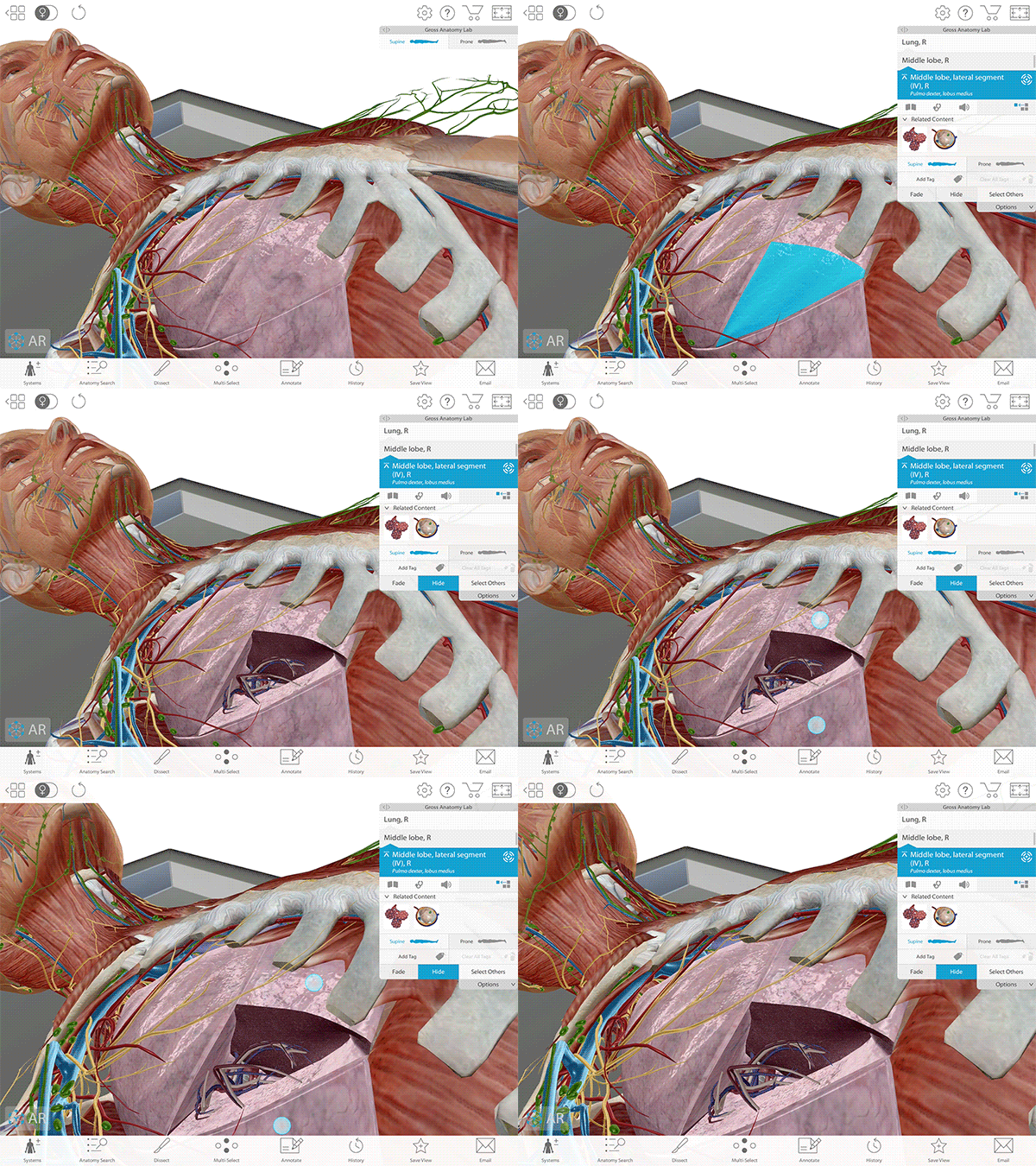
The Gross Anatomy Lab views in Human Anatomy Atlas provide a virtual cadaver lab dissection.
I think that’s the best tool I’ve seen because students can interact with it more and on the same model get to deeper and deeper structures. And I think that conceptual framework is more important before you move into a real person. And seeing it in a real person can sometimes be difficult. It’s real-er. And if you spend enough time you can probably get to the conceptual framework, but you can get lost in the cadaver as well.
So I like the conceptual framework. I think that it’s important, but I really don’t think that it’s a substitute. Now, that said, [...] a cadaver lab, it’s not just ventilation, expenses, and everything else. You need a lot more time. That’s the type of thing where you’re in there 8 hours a day, continually moving through the structures to gain an understanding.
You recently changed from asking students to purchase a published lab manual to using Visible Body and creating your own lab activities. What were the challenges of switching?
The UMass Amherst Science and Engineering Library purchased a Visible Body site license to replace their traditional lab manual.
In the words of librarian Ellen Lutz: "The lab manual that the students usually purchased is $80, and they have around 600 students that come through the A&P class each year, so the math means $48,000—and I can tell you that we do not pay anywhere near $48,000 a year for our site license. As a librarian, that was really what attracted me to it. Our libraries, and our campus in general, are really aware of the rising costs of textbooks and materials that students have to buy. Finding ways that libraries can help support and reduce that burden on students is an important piece of our mission."
Jay explained that he had switched his class to a custom-made set of labs constructed by him and the graduate TAs teaching the lab sections. Jay also mentioned that this transition had its challenges, but is ultimately working out fairly well.
JG: So this last Fall was the first time we had actually gone to “we’ll create our own lab manual,” and it was a rough transition because the TAs hadn’t done it before and [...] I didn’t want to just do everything myself. [This is] partly because I didn’t have time, [and] also because I’m not teaching the labs and if you’re going to be a lab instructor, writing about or creating the lab that you would like the students to do is a big part of it. So I would say last semester was a start of going in the direction we wanted to but it didn’t go that well. In a lot of ways, instructors and students needed to see what didn’t work to know what to correct.
This semester we’ve seen huge improvements. The lab manual’s improved, and now we’re starting to have a really robust lab manual.
One of the things that’s helped a lot, especially in this transition to online learning that we’re doing with the coronavirus, is that the manuals you gave us for Visible Body give us an additional step-by-step process that students can go through with Visible Body to look for certain structures.
Now, some of the structures we don’t use, some of them we do. We look for some structures that aren’t in those labs, but the 3D virtual cadaver and the ability for students to interact with it complements other activities that we do in other formats.
I would say the students have a lot more interaction with the material now than they did before and that’s one of the reasons lab is going so well this semester.
VB: So when you say it went rough the first time, is it because the labs the TAs were writing is what drove that 50-minute session, and so maybe some of them put too much in it or too little or how the students ran through the lab was difficult? What was that challenge exactly?
JG: The challenge was that the TAs (lab instructors) had to create something new and they hadn’t done it before. [They] take responsibility for the curriculum. We divide each lab: each instructor will do a subset of the labs and develop the activities in the lab as well as the preparation. So, having not done that before, students aren’t going to be good at that the first time, but it’s a great experience and they’re a lot more enthusiastic now that it’s going well.
VB: So it’s been an interesting challenge and a fun challenge for those TAs to figure out how to make it work for their students?
JG: Exactly, exactly. And they’re really invested in the student learning as well, so part of that whole transition [was] to create something that’s optimized for our 50-minute labs.
One way students can use Human Anatomy Atlas to study is by annotating the models in 3D.
One of the things that students do in lab now, because they’re prepared, is a snap and chat activity. The iPads have Visible Body, but they have a camera, and so for the list of structures, students come and they will take a picture of each structure and talk about it with the lab instructors or fellow students.
If they’re confused, they have their manual, which is Visible Body. [They can ask] “Where is this structure? How am I gonna find it? What model will I find it on?” and they’ll go in and find it and they’re able to do that pretty quickly.
Students can bring models from Human Anatomy Atlas into the lab (or anywhere, really) on compatible devices with Atlas’ augmented reality (AR) feature.
Your school, like so many others, is continuing the semester online. How will that change your course?
JG: I stream our class, and our online response system and lecture is on the internet. I’ve had as many as a hundred people regularly attending virtually, so it doesn’t change lecture because [...] we have a lot of tools through our textbook but also Visible Body.
What we’ve done is added more Visible Body to remote labs. The students are already well-versed in it, so it’s not going to be a transition. A lot of the solutions are easy, so it’s been nice to at least tell people what I do.
We would like to thank Jay very much for his time and ideas—seriously, he’s a busy person and we really appreciate it!—and we hope that his strategies for incorporating 3D technology into his lab and lecture have inspired you! Happy teaching!
Also, you can check out Visible Body’s collection of lab manuals (for the A&P, Atlas, and Physiology & Pathology apps) here!
Be sure to subscribe to the Visible Body Blog for more anatomy awesomeness!
Are you an instructor? We have award-winning 3D products and resources for your anatomy and physiology course! Learn more here.


