

New Content Added to Human Anatomy Atlas 2022 +!
Here at Visible Body, we’re always looking for ways to improve and expand our interactive 3D content library. After listening to instructors and students who use Visible Body products to teach and learn, we've launched new and updated anatomy content in Human Anatomy Atlas 2022 +!
Today on the blog, we’ll check out the new and updated models available now on a computer or mobile device near you!
New models
Heart wall
The heart wall model, originally created for Physiology & Pathology, shows a detailed view of the layers of the heart wall.
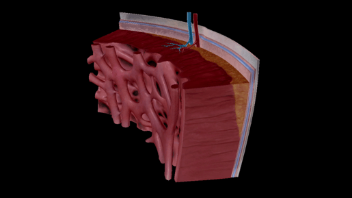
Image from Human Anatomy Atlas 2022 +.
The heart wall is made up of three layers: the epicardium, the myocardium, and endocardium.
The epicardium is the outer layer of the heart. The epicardium is made up of the visceral pericardium (the pericardium is a fluid-filled sac that surrounds the heart), connective tissue, and adipose tissue. These tissues cushion the heart and bind the epicardium to the myocardium.
The myocardium is the middle layer of the heart. It’s made up of cardiac muscle cells (cardiomyocytes), and it’s the thickest layer of the heart wall. The myocardium is responsible for the heart’s contractions.
The endocardium is the innermost layer. It’s actually made up of two layers: the outer layer is made of connective tissue that connects to the myocardium, and the innermost layer is made up of endothelial cells that make a nonstick surface for blood flow. The endocardium covers the inner surfaces of the heart, including heart valves.
Skeletal muscle
The skeletal muscle model is one of our most highly-requested models! It shows the macro- and microelements of skeletal muscle from bone attachment to the makeup of the individual muscle fibers.
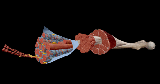
Image from Human Anatomy Atlas 2022 +.
Skeletal muscle contains a lot of layers and a lot of connective tissue, so let’s dive right in.
Each muscle fiber consists of myofibrils, rod-shaped fibers that contain thousands of sarcomeres, the smallest functional unit of skeletal muscle fibers. Sarcomeres contain contractile, regulatory, and structural proteins. The shortening of sarcomeres leads to the shortening of the whole muscle.
Muscle fibers are surrounded by a connective tissue layer made of collagen and reticular fibers; this is called the endomysium. The endomysium transfers the force of the muscle fibers to the tendons. The collagen in all of the connective tissue layers intertwines with the tendon’s collagen.
Fascicles are bundles of muscle fibers and are surrounded by the perimysium, a middle layer of connective tissue. The epimysium separates muscles from other tissues and organs and allows the muscle to retain its structure and move independently.
In addition to movement, skeletal muscle helps support the body, maintains its posture, stores amino acids, and helps maintain thermostasis. In the case of starvation, skeletal muscle can even be used as an energy source.
Hepatic lobule
The hepatic lobule model is a close-up view of the functional unit of the liver, showcasing hepatocytes, sinusoids, and more.
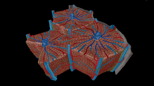
Image from Human Anatomy Atlas 2022 +.
The hepatic lobule consists of hepatocytes collected in a hexagonal shape around a central vein. Hepatocytes comprise eighty percent of the liver’s volume! Hepatocytes perform several functions, including the formation and secretion of bile, storing glycogen, detoxification, metabolization of cholesterol and fat, and synthesizing urea.
Between the cords of hepatocytes is a vascular space that contains an endothelium and a sinusoid. The sinusoids contain Kupffer cells, which destroy bacteria, and stellate cells, which respond to injury in the liver and store vitamin A.
Grooves in the cell membranes form bile canaliculi, small ducts from which bile flows. These canaliculi merge to form the right and left hepatic ducts, which merge to form the common hepatic duct.
Updated models
Osteon
We’ve completely revamped our osteon model to show more detail of the central canal with its surrounding concentric lamellae.
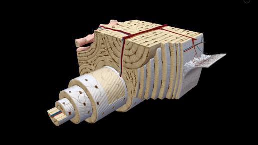
Image from Human Anatomy Atlas 2022 +.
Osteons are structural units of compact bone. Each osteon consists of a central canal, which contains nerve filaments and one or two blood vessels, surrounded by lamellae. Lamellae are concentric rings of bone matrix made up of osteocytes. Lacunae, small chambers containing osteocytes, are arranged concentrically around the central or Haversian canal.
Together, the central canal and the lamellae make up an osteon, the basic unit of structure of compact bone.
Sectioned femur
The updated sectioned femur model shows more regions of compact and spongy bone, along with a section of periosteum and blood supply to the bone.
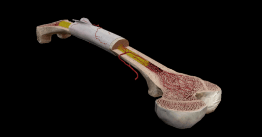
Image from Human Anatomy Atlas 2022 +.
The femur is the longest bone in the human body and is also one of the strongest—after all, the femur supports your entire upper body. The femur contains a marrow-filled medullary cavity, which is present in all of the long bones of the limbs.
Bone marrow fills the cavities of long bones and occupies the spaces of spongy bone. Yellow marrow, consisting mostly of fat, is found in the central cavities of long bones. Red marrow is found in the medullary cavities of flat and short bones, articular ends of long bones, vertebral bodies, spongy bone of the cranium, sternum, ribs, and scapulae.
The periosteum is the membrane that covers the surfaces of bones. It consists of two layers: an outer fibrous layer and an inner osteoblastic layer. The outer layer is mostly made up of collagen, and it also contains blood vessels that supply blood to the osteocytes. The inner layer contains osteoblasts, cells that produce bone. Osteoblasts are more prominent in fetal development and early childhood, but in the adult body, they help remodel bone.
Wrap up
Thanks for coming along on this tour of the new and updated models now available in Human Anatomy Atlas 2022 +! Click here to learn more.
If you want to read more about relevant anatomy, check out these blog posts:
- 3D Skeletal System: Compact Bone, Spongy Bone, and Osteons—Oh My!
- The Toxic Substance Treatment Plant: Liver Anatomy
- That Boom Boom Pow: Virtual Dissection (sort of) of the Human Heart
Be sure to subscribe to the Visible Body Blog for more anatomy awesomeness!
Are you an instructor? We have award-winning 3D products and resources for your anatomy and physiology course! Learn more here.
