Posted on 1/22/13 by Maite Suarez-Rivas
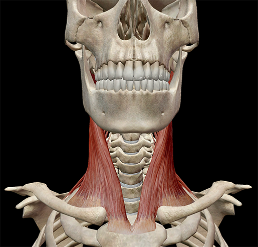 Image from Muscle Premium.
You know what I love about my neck muscles? EVERYTHING. They allow me to move my head so I can do all sorts of things, such as look at the Chipotle menu, eat fro-yo, or turn to look at my coworker (who has so thoughtfully just brought me a piece of cake). Okay, basically they let me stuff my face, but the neck muscles do much more than that! The neck muscles (and neck anatomy on the whole) are responsible for head movement, stabilizing the upper region of the body, assisting in swallowing, helping to elevate the rib cage during inhalation, and more.
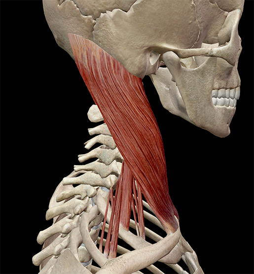
Image from Muscle Premium.
If you press against your neck, you'll feel muscles (obviously). However, the neck region is home to more than just neck muscles; muscles of the head, spine, and thorax also attach in this area.
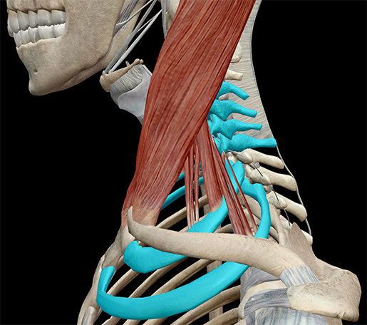 Image from Muscle Premium.
The neck muscles include the scalenes, which attach the cervical vertebrae to the thoracic cage, and the sternocleidomastoid, which attaches the skull to the thoracic cage. These muscles move the head and neck.
There are also muscles that act on the hyoid and laryngeal skeleton (sternohyoid, sternothyroid, thyrohyoid, omohyoid, stylohyoid) and the mandible (mylohyoid and digastric).
I mentioned the scalenes above. Let's take a look at them! As always, if you have Muscle Premium, feel free to follow along!
The scalenes are a series of muscles that work like scaffolding, connecting the cervical vertebrae to the thoracic cage. The scalenes originate on the transverse processes of the cervical vertebrae (C02–C07) and attach to the first and second ribs of the thoracic cage.
|
|
Origin |
Insertion |
|
Scalenus anterior |
Anterior tubercles of the transverse processes of C03-C06 |
Scalene tubercle on the inner border and upper surface of the first rib |
|
Scalenus medius |
Posterior tubercles of the transverse processes of C02-C07 |
Upper surface of the first rib |
|
Scalenus posterior |
Posterior tubercles of the transverse processes of C05-C07 |
Outer surface of the second rib; occasionally blended with the scalenus medius |
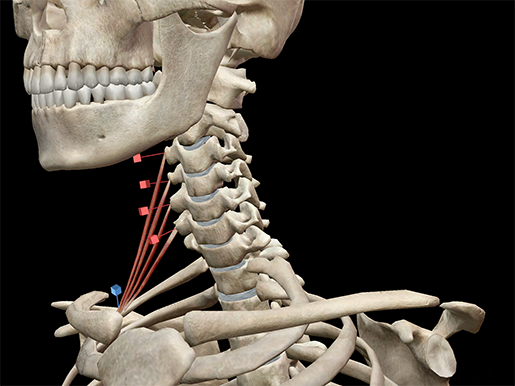 Scalenus anterior. Image from Muscle Premium.
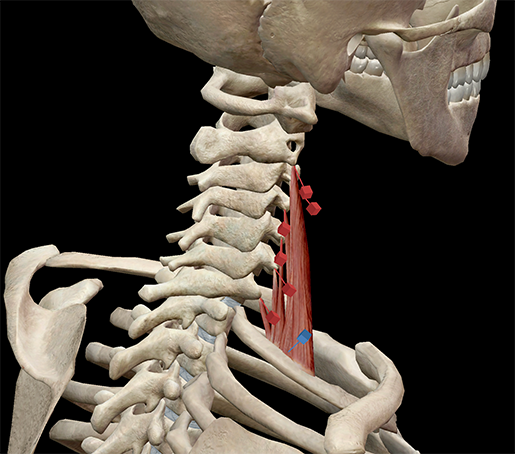
Scalenus medius. Image from Muscle Premium.
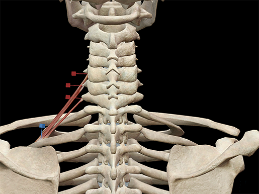
Scalenus posterior. Image from Muscle Premium.
Watch the scalenes work to support lateral flexion of the neck.
Video footage from Muscle Premium.
Neck pain can be a, well, real pain in the neck! Neck pain is a very common ailment, and its causes are many—anything from a car accident, to poor posture, to simply sleeping the wrong way.
Muscle strains are a common cause. Overuse of the neck muscles, such as excessive exercise or staying in an uncomfortable position too long (hunched over your computer, for example), can cause strains. Even grinding your teeth at night can do it. Wear and tear on the neck over time can cause pain, including forms of arthritis.
Most neck pain responds well to over-the-counter anti-inflammatories, such as Tylenol or Ibuprofen. Rest and alternating heat/cold compresses can help ease pain. Gentle stretching, such as stretching in to the pain, can help alleviate it.
Be sure to subscribe to the Visible Body Blog for more anatomy awesomeness!
Are you an instructor? We have award-winning 3D products and resources for your anatomy and physiology course! Learn more here.
- Learn Muscle Anatomy: Muscles of Mastication
- Learn Muscle Anatomy: Knee Joint Group
- Learn Muscle Anatomy: Gastrocnemius
Additional Sources:
When you select "Subscribe" you will start receiving our email newsletter. Use the links at the bottom of any email to manage the type of emails you receive or to unsubscribe. See our privacy policy for additional details.
©2025 Visible Body, a division of Cengage Learning