Posted on 1/11/13 by Courtney Smith
Have you ever made faces at yourself in the mirror? What am I talking about, of course you have (when you're brushing your teeth, watch the hilarious things your eyebrows do).
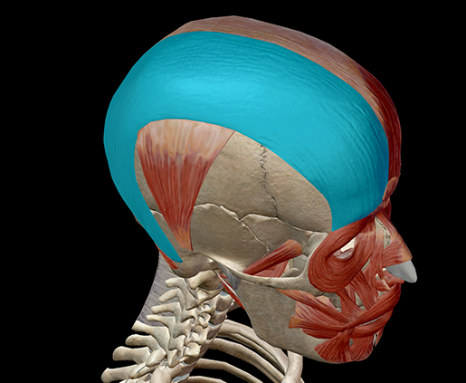
Many of the expressive things we do with our face are thanks to the facial muscles of expression. If you've a mirror handy, take a look in it and furrow your forehead or lift your eyebrows. One of the largest of the expression muscles is responsible for those faces you're making: the occipitofrontalis.

Image from Human Anatomy Atlas.
The occipitofrontalis is an interesting muscle. It is comprised of three sections: the frontalis, the occipitalis, and the galea aponeurotica. Each section is responsible for a different action involving the scalp, forehead, or brows.
Let's take a look, shall we?
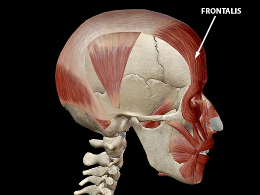
The frontalis muscle is a thin, flat band in the anterior scalp. It has several origin points: the procerus muscle, the corrugator supercilii muscle, the orbicularis oculi fascia, and the zygomatic process of the frontal bone. It obviously covers a lot of ground. It inserts on the superficial fascia of the facial muscles, as well as the skin above the nose and eyes.
 Image from Human Anatomy Atlas.
The frontalis muscle is at work when the scalp is drawn anteriorly—for example, when you lift your eyebrows, which also lifts the skin above your eyes and nose. Press your fingers to the bridge of your nose and waggle your eyebrows; feel how the muscle pulls at the skin?
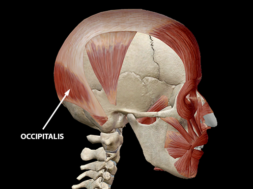
The occipitalis is a thin quadrilateral muscle in the posterior scalp. It originates on the occipital bone and the mastoid process of the temporal bone. It inserts into the galea aponeurotica.
The occipitalis draws the scalp posteriorly.
 Image from Human Anatomy Atlas.
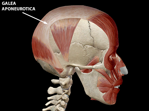
The galea aponeurotica (which is hella fun to say) is a broad, fibrous sheet covering the upper part of the cranium. It originates on the occipital bone and inserts into both the occipitalis and frontalis muscles.
 Image from Human Anatomy Atlas.
The aponeurosis helps with scalp movement.
Pain in the occipitofrontalis is usually found in either the frontalis or the occipitalis. In the frontalis, the pain is usually in the forehead; in the occipitalis, there can be moderate pain at the back of the skull, which sometimes refers to the suboccipital and causes severe pain at the back of the eye. This can sometimes lead one to believe they are having a stroke (yikes!).
Be sure to subscribe to the Visible Body Blog for more anatomy awesomeness!
Are you a professor (or know someone who is)? We have awesome visuals and resources for your anatomy and physiology course! Learn more here.
Additional Sources:
- American Academy of Manual Medicine
- NBCI
When you select "Subscribe" you will start receiving our email newsletter. Use the links at the bottom of any email to manage the type of emails you receive or to unsubscribe. See our privacy policy for additional details.
©2025 Visible Body, a division of Cengage Learning