Posted on 1/2/13 by Courtney Smith
It's kind of serendipitous that I start writing a thing about the lateral rotator muscles when a Lewis Black schtick comes on my iTunes about mixing up FEMA and femur.
The lateral rotators are muscles in the hip/gluteal region of the body and their main job is basically what it sounds like: to rotate the hip joint laterally. To a lesser extent, they also help with other motions of the hip, such as extension and adduction.
Want to learn more about the lateral rotators of the hip? Check out our free eBook!
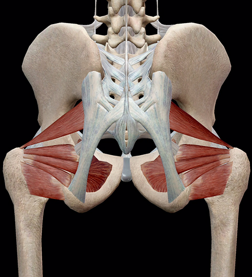
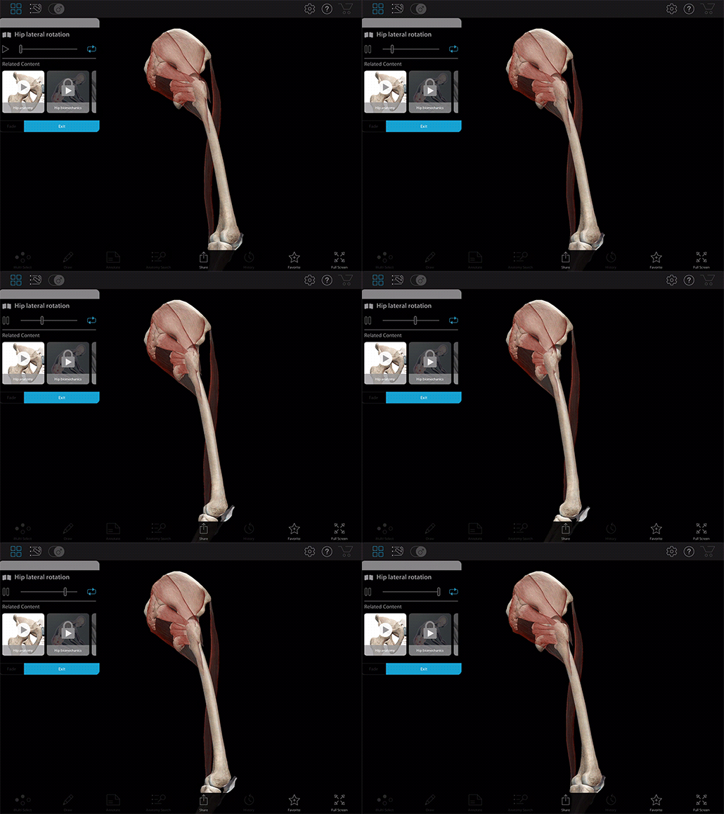
The lateral rotators in context and in action. Images and video footage from Muscle Premium.
The lateral rotators are: the superior gemellus, inferior gemellus, obturator externus, obturator internus, quadratus femoris, and the piriformis. These muscles all originate on the pelvic area and insert onto the greater trochanter of the femur.
Let's take a quick dive into each of them, shall we?
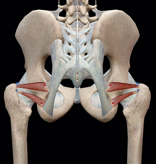
Image from Muscle Premium.
The superior gemellus and inferior gemellus are long, thin muscles that originate on the ischium and insert on the medial surface of the greater trochanter.
Both muscles, in addition to helping rotate the hip joint laterally, help to abduct the flexed thigh.
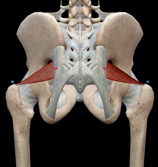
Image from Muscle Premium.
The piriformis is a long muscle that originates on the anterior surface of the lateral process of the sacrum and gluteal surface of the ilium at the margin of the greater sciatic notch. It inserts on the superior border of the greater trochanter.
Like the gemellus muscles, the piriformis rotates the hip laterally. If the thigh is flexed, it also helps to abduct it.
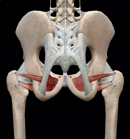
Image from Muscle Premium.
I love these two. They have such interesting shapes—they kind of look like comic book word bubbles if you turn your head and squint.
The externus originates on the external surface of the obturator membrane and the margins of obturator foramen (the hole created by the pubis and ischium); the internus originates on the inner surface of the obturator foramen and the surfaces of the surrounding bones. They insert onto the posteromedial surface of the greater trochanter.
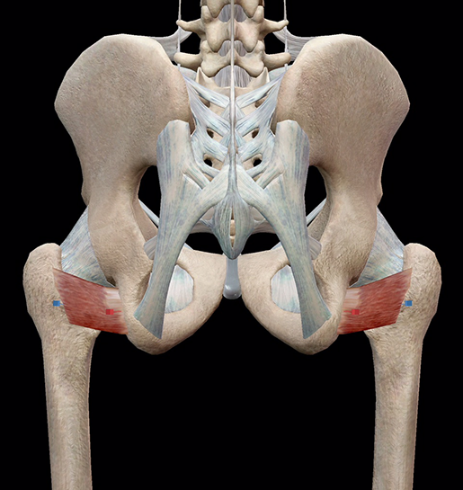
Image from Muscle Premium.
The quadratus femoris is a short, strong, rectangular muscle that originates on the ischial tuberosity and inserts onto the intertrochanteric crest.
That was kind of a lot of information to throw at you. Let's recap:
|
Muscle |
Origin |
Insertion |
Action |
|
Superior gemellus |
Ischium |
Medial surface of the greater trochanter |
Laterally rotates hip joint |
|
Inferior gemellus |
Ischium |
Medial surface of the greater trochanter |
Laterally rotates hip joint |
|
Piriformis |
Anterior surface of the lateral process of the sacrum and the gluteal surface of the ilium at the margin of the greater sciatic notch |
Superior border of the greater trochanter |
Laterally rotates hip joint; abducts thigh when flexed |
|
Obturator externus |
External surface of the obturator membrane and the margins of obturator foramen |
Trochanteric fossa |
Laterally rotates hip joint; abducts thigh when flexed |
|
Obturator internus |
Internal surface of the obturator foramen and the surfaces of the surrounding bones |
Posteromedial surface of the greater trochanter |
Laterally rotates hip joint; abducts thigh when flexed |
|
Quadratus femoris |
Ischial tuberosity |
Intertrochanteric crest |
Externally rotates and adducts the hip joint |
Be sure to subscribe to the Visible Body Blog for more anatomy awesomeness!
Are you an instructor? We have award-winning 3D products and resources for your anatomy and physiology course! Learn more here.
Additional Sources:
When you select "Subscribe" you will start receiving our email newsletter. Use the links at the bottom of any email to manage the type of emails you receive or to unsubscribe. See our privacy policy for additional details.
©2025 Visible Body, a division of Cengage Learning