Learn Heart Anatomy: Vessels, Valves, and Chambers (oh my!)
Posted on 12/10/12 by Courtney Smith
Your heart is an extraordinary machine.
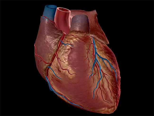
Have you ever seen the PIXAR movie Wall-E? In the movie, every robot has a directive, or purpose, that it must carry out without fail, because it's an ingrained part of their programming. Think of your heart in those terms: your heart has one directive, which is to pump blood. That's it. Other organs can have more than one purpose, but the heart does this one, single thing. If it fails to do so, everything breaks down.
 Image captured from Human Anatomy Atlas.
My obvious love for all things PIXAR aside, my point still stands. The heart is an organic machine that allows you to operate at maximum capacity. Let's take a look at the various anatomy of the heart and how they keep the cogs moving, so to speak.
Want more heart facts? Check out our free "Build a Heart" eBook!
The Heart Chambers
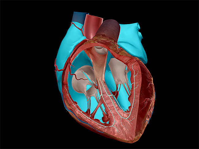
The most basic component of the heart is its role as a four-chambered muscle. The heart wall is composed of three distinct layers: the epicardium (outermost layer), the myocardium (middle layer, comprised of muscular fibers), and the endocardium (innermost layer). The heart pumps due to the contractions of the muscles in the myocardium; the muscle contractions are triggered by the electrical impulses delivered by the conduction system.
 Image captured from Human Anatomy Atlas.
The four chambers of the heart allow blood to pump in and out to specific locations.
|
Chamber |
Role |
|
Left atrium |
Receives oxygenated blood from pulmonary veins and empties it into the left ventricle. |
|
Right atrium |
Receives deoxygenated blood from the vena cavae and coronary sinus and empties it into the right ventricle. |
|
Left ventricle |
Pumps oxygenated blood into the aorta via the tricuspid aortic valve; also forms the apex of the heart. |
|
Right ventricle |
Pumps deoxygenated blood into the pulmonary trunk via the pulmonary valve. |
To pump blood in and out of the chambers, doors are needed. The heart's valves openand shut, regulating the amount of blood that enters and exits. The atrioventricular valves (the tricuspid and mitral valves) control blood flow into the ventricles. They are pulled open by fibrous cords called chordae tendineae. The pulmonary and aortic valves control the flow of blood out of the heart; the pulmonary valve controls the flow of blood into the pulmonary trunk, and the aortic valve into the aorta.
The Great Vessels of the Heart
Pumping blood out of the heart would be a messy affair if not for the sprawling, closed-loop circuit of vasculature in the body. The great vessels of the heart provide passage of blood to and from the arteries and veins. There are four great vessels in all (corresponding to the number of chambers and valves).
|
Vessel |
Role |
|
Aorta |
Largest artery in the body; receives blood from the left ventricle via the aortic valve; branches into the left common carotid, left subclavian, and brachiocephalic trunk. |
|
Pulmonary trunk |
Artery that supports pulmonary circulation by carrying deoxygenated blood from the right ventricle to the lungs for gas exchange; the pulmonary trunk and its branching arteries are the only arteries in the body that carry deoxygenated blood. |
|
Superior vena cava |
Vein that drains deoxygenated blood from the upper half of the body, which is received by the right atrium. |
|
Inferior vena cava |
Vein that brings deoxygenated blood from the lower half of the body, which is received by the right atrium. |
The Coronary Vessels of the Heart
Not only does the heart supply the body with blood, but it also supplies blood to itself. The coronary vessels provide the heart tissue with blood; the coronary arteries supply blood to the heart tissue while the veins move oxygenated blood from the heart tissue into the right atrium.
A myocardial infarction, or heart attack, occurs when blood flow to the heart tissue via the coronary arteries is obstructed (usually due to plaque buildup). If oxygenated blood can't reach the tissue, the tissue will die, weakening the heart and leaving behind scarring.
If an artery is blocked, it may warrant coronary bypass surgery, in which a section of a healthy vessel is used to create a new pathway, detouring blood from the blocked artery into the new vessel, allowing oxygenated blood to flow.
The Conduction System of the Heart
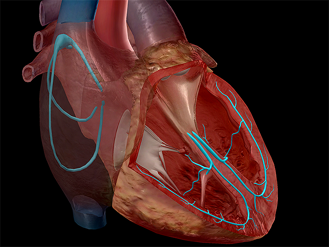
Have you ever wondered why your heart continues to beat without fail? It's all thanks to the conduction system. The conduction system is a system controlled by the autonomic nervous system, which delivers electrical impulses that cause the cardiac muscles to contract, or beat. Each contraction pumps blood throughout the cardiovascular system.
 Image captured from Human Anatomy Atlas.
The conduction system is comprised of a series of nodes and fiber bundles, and each electrical impulse travels the same path. The sinoatrial node is where the impulse initiates and then travels to the atrioventricular node (where it pauses), then further to the bundle of His. Between the sinoatrial and atrioventricular nodes, the atria contract, pumping blood into the ventricles. From the bundle of His, the impulse travels through the bundle branches of each ventricle, then culminates at the Purkinje fibers. Between the bundles branches and the Purkinje fibers, the ventricles contract, pumping blood out of the heart.
Each electrical impulse takes approximately 0.22 seconds to complete the cycle. On average, the heart beats approximately 100,000 times every day. During the average human lifetime, the heart will beat more than 2.5 billion times! Now that is what I call one well-made machine.
Be sure to subscribe to the Visible Body Blog for more anatomy awesomeness!
Are you a professor (or know someone who is)? We have awesome visuals and resources for your anatomy and physiology course! Learn more here.


