Posted on 10/14/14 by Maite Suarez-Rivas
Twenty-two bones come together like a puzzle to make the skull. Some bones give shape to the face, others protect the brain. But it's not all bones! The skull also includes cartilage (put your finger on the tip of your nose and wiggle it) and ligaments (open and close your mouth if you want to use them).
Here are some other fast facts about the skull.
Want even more facts about the skull? Check out our free Skull Bones eBook!
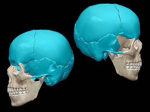
The bones that enclose and protect your brain (like a braincase!) form the neurocranium. Need a list of those bones? Here it is: The bones of the neurocranium are the ethmoid, sphenoid, frontal, and occipital bones (one each), and then there are the parietals and temporals (two each). If you take the ethmoid and the sphenoid out of that list you have the bones of the calvaria (the skullcap). The calvaria is a subdivision of the neurocranium. When you talk about the calvaria, you are talking just about the bones on the superior part of the cranium.
 The calvaria (right) is a subdivision of the neurocranium (left). Images from Human Anatomy Atlas.
Fourteen bones form the facial skeleton. There is the mandible (jaw bone) the vomer (gives shape to your nose) and then a series of paired bones (as in there is a left and a right): the nasals, maxillae, lacrimals, zygomatics, palatines, and the inferior nasal conchae. The face is also formed by the nasal cartilages (a group of connective tissue structures that give shape to the framework of your nose) and by the teeth (the upper arch of teeth are attached to the maxillae and the lower arch of teeth are attached to the mandible).
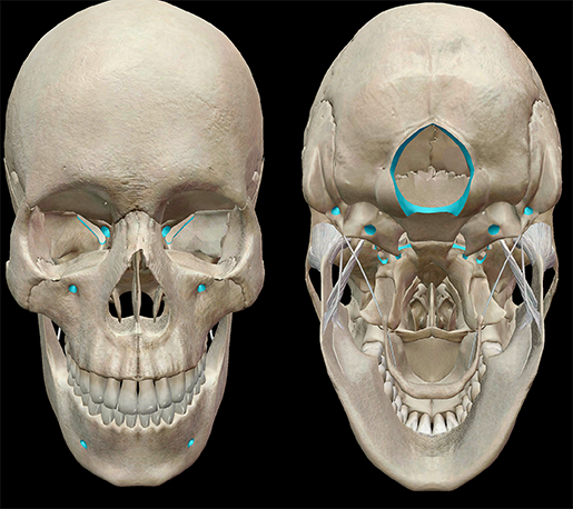
Foramina are apertures, sometimes called canals, scattered throughout the bones of the skull. These openings commonly function as passageways for nerves and vessels. At the base of the skull, in the occipital bone, is the largest foramen of the skull, the Foramen magnum. Vertebral arteries and the spinal cord pass through this opening.

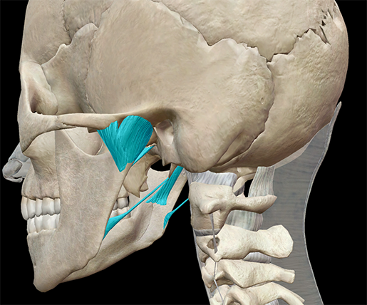
Touch the area of your face right in front of your ear. Now open and close your mouth and you will feel your mandible moving. Each temporomandibular joint is formed by the temporal bone (r,l), the mandible, and ligaments that surround the joint. These ligaments reinforce the area where the cranium articulates with the mandible.

Image from Human Anatomy Atlas.
A number of ligaments reinforce the temporomandibular joint. The main "hinge" of the temporomandibular joint is the sphenomandibular ligament—a flat, thin band that connects the sphenoid bone to the lingula of the mandibular foramen.
The ethmoid bone, located at the roof of the nose and between the eyes, has tiny foramina. Nerve cells in the nose detect odors, carry those signals through the foramina in the ethmoid, and to the olfactory bulbs. From there, the signals move along the olfactory tracts to the brain. The palatine bones help give shape to the back of the roof of the mouth, the floor of the nasal cavity, and the floor of the orbits.
| Did you know? The skull is part of the axial skeleton, which also includes the laryngeal skeleton, vertebral column, and thoracic cage. |
Be sure to subscribe to the Visible Body Blog for more anatomy awesomeness!
Are you an instructor? We have award-winning 3D products and resources for your anatomy and physiology course! Learn more here.
Additional Sources:
When you select "Subscribe" you will start receiving our email newsletter. Use the links at the bottom of any email to manage the type of emails you receive or to unsubscribe. See our privacy policy for additional details.
©2025 Visible Body. All Rights Reserved.