Anatomy and Physiology: Internal Female Reproductive Anatomy
Posted on 4/26/13 by Courtney Smith
%20(2).jpg?width=515&height=386&name=screenshot%20(6)%20(2).jpg)
Image from Visible Body Suite.
Ah, the uterus, nature's Rubix cube. You're probably laughing to yourself, saying, "It's not some great mystery—I know enough about this to get by." Do you? Do you really? I'm not asking to be mean. It may come as a surprise, but most people don't know much about reproductive anatomy—even their own!
This is the first of two posts exploring the internal reproductive systems. Let's take a look at the female system first.
Fallopian, say what?
%20(1).jpg?width=515&height=386&name=screenshot%20(7)%20(1).jpg)
Image from Visible Body Suite.
Once upon a time when I had sex ed in the 7th grade (for, like, one day), my health teacher tried to describe the internal reproductive system to us. He told us to picture a kung-fu crane stance, with the uterus as the kung-fu master and the fallopian tubes as his arms (like this). Ridiculous as that sounds, none of us ever forgot it.
The fallopian tube, or oviduct, is about 10 cm long and consists of three coats: serous, muscular, and mucous. Each fallopian tube consists of three portions: the isthmus (a constricted section connected to the uterus), ampulla (intermediate dilated portion that curves over the ovary), and infundibulum (open to the abdomen).
Ova (eggs) travel through the fallopian tube to the uterine cavity.
Fimbriae, or that thing you probably have never heard of
%20(1).jpg?width=515&height=386&name=screenshot%20(8)%20(1).jpg)
Image from Visible Body Suite.
Fimbriae are interesting structures. Remember my health teacher's crane stance? Think of the fimbriae as the hands of the kung-fu master. FYI, I'm going to run this analogy into the ground.
Fingerlike in shape, the fimbriae are projections that are attached to the ovaries. Before ovulation (the release of an ovum), the fimbriae become positioned close to the ovarian follicle surface. Cilia, hairlike organelles, on the fimbriae beat in rhythmic waves, creating currents that sweep the ovum into the fallopian tube.
Ovaries, and the stuff inside them
%20(1).jpg?width=515&height=386&name=screenshot%20(9)%20(1).jpg)
Image from Visible Body Suite.
Okay, so if the fallopian tubes are the arms of the kung-fu crane stance and the fimbriae are the hands, then the ovaries are nunchucks. The ovaries are gonads (which are derived from the same set in utero), which produce gametes (ova). These gametes contribute half the genetic material needed for embryonic development (the other half comes from the male gametes).
Ovaries are endocrine glands, producing the hormones progesterone and estrogen—sex hormones that govern early development and contribute to the menstrual cycle. Each ovary is about 4 cm long and 2 cm wide. The ovarian ligament attaches it to the uterus while a suspensory ligament attaches it to the pelvic wall.
During ovulation, an ovum is dispensed from an ovary's surface and is carried into the fallopian tube, where it proceeds to the uterus.
What is the uterus?
%20(1).jpg?width=515&height=386&name=screenshot%20(10)%20(1).jpg)
Image from Visible Body Suite.
If I went up to you right now and asked you to say what the uterus is and does, could you? Let's try it. Without opening up Google or Wikipedia, can you give a clear, concise explanation of the uterus? "That thing where babies happen" doesn't cut it. See? It's tough to know what you're never really taught.
The uterus is a hollow, thick-walled, pear-shaped organ—a duct, actually!—located in the pelvic cavity between the bladder and the rectum. The fallopian tubes lead into the upper part of the uterus, one on either side, while the lower part of the uterus leads to the vagina. Ligaments hold the upper part of the uterus in suspension; the lower part is embedded in fibrous tissue.
That's all well and good, I can hear some of you say, but what does it do? Well, when an ovum (egg) is fertilized, it embeds itself into the uterine wall and is normally retained there as it develops. Development of an embryo and then a fetus causes the uterine wall to expand in size—enormously, actually. You've seen pregnant people. You know.
After labor, the uterus shrinks down to almost its normal size (crazy, right?), although its cavity is larger and the muscular layers are more defined (because it just pushed out a baby, like a boss).
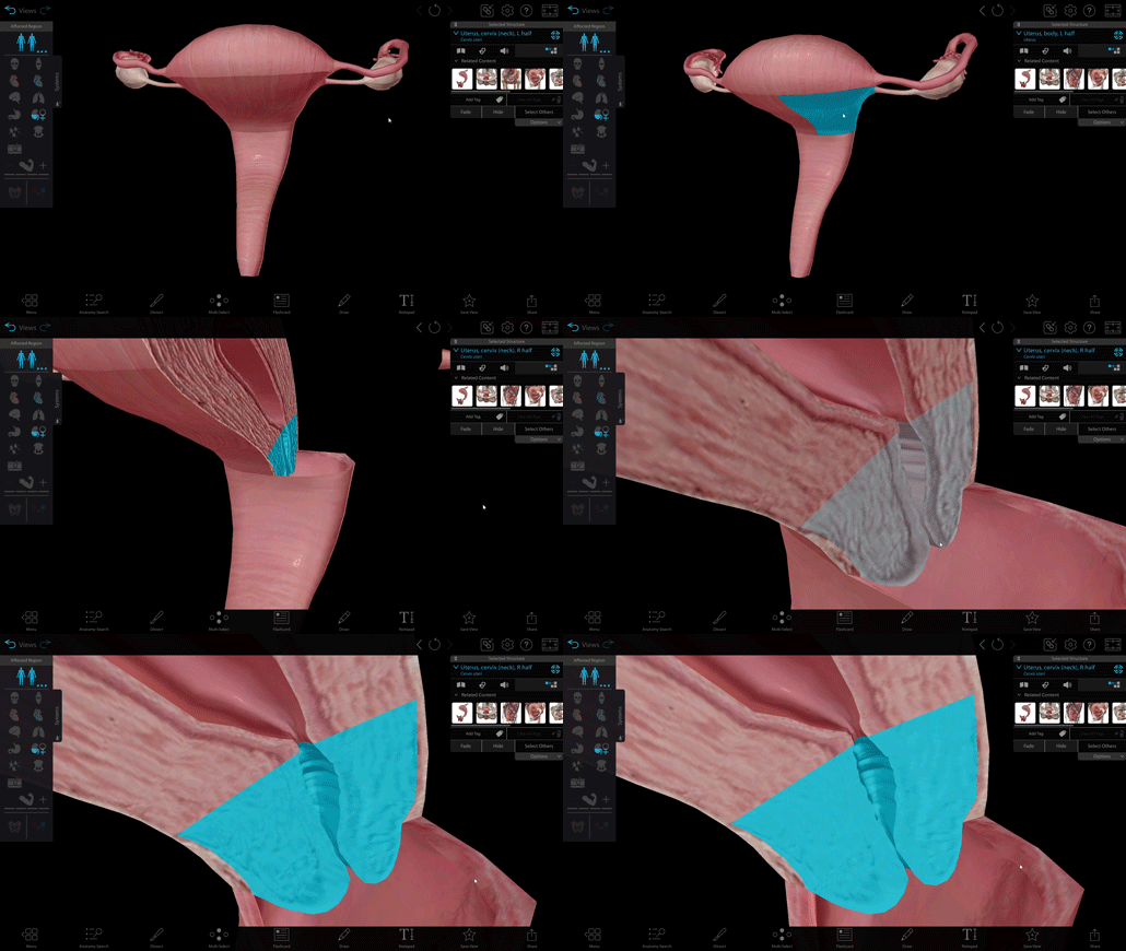
The cervix

GIF from Visible Body Suite.
On one memorable occasion, some friends of mine and I were discussing what we knew about each other's anatomy over a plate of potato skins (I have interesting friends and conversations, sometimes), when one of my guy friends shouted triumphantly, "Hah! The cervix! It's that … that thing.…" Yes, indeed, friend. The cervix is that thing.
The cervix is actually the lowest part of the uterus, a constricted, somewhat conically shaped segment that leads to the vagina. It is the passageway for menstrual flow, entering sperm, and childbirth (it's a trifecta of hilarity and terror). It also secretes a clear alkaline mucus that changes character depending on where someone is in their menstrual cycle.
And there you have it! The basic rundown of everything you always wanted to know about the internal female reproductive system. Stay tuned for our next post, where we'll tackle the male system.
Do you have Visible Body Suite and want to keep the learning going? Study the structures of the female reproductive system with our premade Flashcard deck! Here's how to open them in VB SUite:
- Copy this link:
https://apps.visiblebody.com/share/?p=vbhaa&t=4_55692_637636798136890000_566932 - Use the Share Link button in VB Suite.
- Paste the link to view the deck.
- Save the deck to your User Account.
For more premade Flashcard decks on popular topics, check out our whole library here.
Be sure to subscribe to the Visible Body Blog for more anatomy awesomeness!
Are you an instructor? We have award-winning 3D products and resources for your anatomy and physiology course! Learn more here.
Related Posts:
- Pregnant Pause: A look at ovulation, fertilization, and implantation
- Anatomy and Physiology: Homologues of Reproductive Anatomy
- Anatomy and Physiology: Internal Male Reproductive Anatomy


