Anatomy and Physiology of the Lower Respiratory System
Posted on 3/23/18 by Katrina Irwin
We breathe anywhere from 17,000 to 24,000 times a day (or more!), and most of the time we don't even think about it. We can focus on the more important things, like how the lower respiratory system works. Let’s take a look!
Trachea
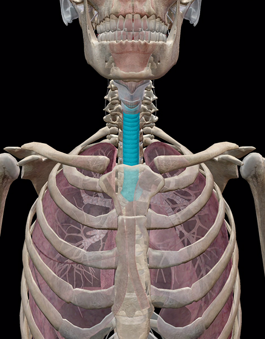 Image from Human Anatomy Atlas.
The trachea, also known as the windpipe, is the major airway of the lower respiratory system. It is a membrane made of cartilage that starts at the larynx and continues to the fifth thoracic vertebrae. The trachea is about eleven centimeters long and 2.5 centimeters wide. Wrapped around it are irregular rings composed of hyaline cartilage and elastic fibrous membranes with mucus interior linings, which help provide structure to keep the airway open. There are usually 16 to 20 of them! At the fifth vertebrae, the trachea divides into two bronchi, but...before we dig deeper let's take a look at the lungs themselves.
The Left and Right Lungs
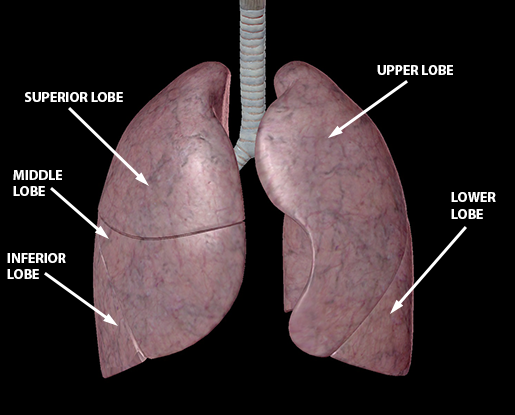 Image from Human Anatomy Atlas.
We all know that we have two lungs, but the left and right lungs are structurally different from each other. The left lung is divided into two lobes, an upper and a lower, and the right lung is divided into three lobes, the superior, the middle, and the inferior. The lungs are made of a porous spongy texture and are highly elastic. A delicate double layer serous membrane called the pleura covers the surface of the lung and dips into the fissures between the lobes. There is a fluid between the fissures to prevent the lungs from moving and chafing against the thorax wall. The left lung has a indentation (cardiac notch) to accommodate the heart. The main function of the lungs is to provide oxygen to capillaries so they can oxygenate the blood.
Fun fact: The total surface area of the lungs varies from 50 to 75 square meters (540 to 810 sq ft)—about the same area as one side of a tennis court!
Primary Bronchus
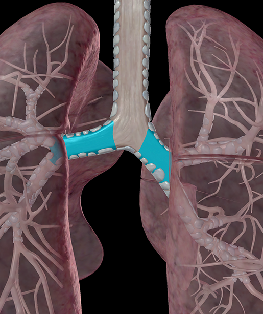 Image from Human Anatomy Atlas.
The primary bronchi link the trachea to the left and right lungs. Similar to the trachea, they are composed of incomplete rings of hyaline cartilage, with the right bronchus membrane containing six to eight cartilaginous rings, and the left with nine to 12. The right bronchus is typically wider and shorter, and is more vertical than the left. Due to this, inhaled objects tend to lodge themselves more in the right bronchi than the left.
Due to the asymmetry of the lungs, the bronchi split into a different number of lobes.The right bronchus divides into three major secondary bronchi that lead to the three main lobes of the right lung. The left primary bronchus bifurcates into two secondary bronchi that then service the two lobes of the left lung.
Intermediate Bronchus
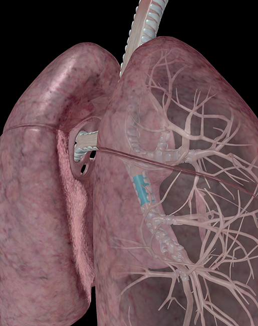 Image from Human Anatomy Atlas.
There are many names for these bronchi. The intermediate bronchus is also called the secondary bronchus, lobar bronchus, or intrapulmonary bronchus. This is the airway of the lower respiratory system. These bronchi branch from the primary bronchi and service the lobes of the left and right lungs. Within the lung itself, the lobar bronchi divide and subdivide throughout the lungs with the smallest subdivisions forming tubes called bronchioles. Instead of rings, the secondary bronchi are supported by irregular plates of hyaline cartilage. Branch by branch, the bronchi become smaller airways, delivering oxygen-rich air to the lungs.
Tertiary (Segmental) Bronchus
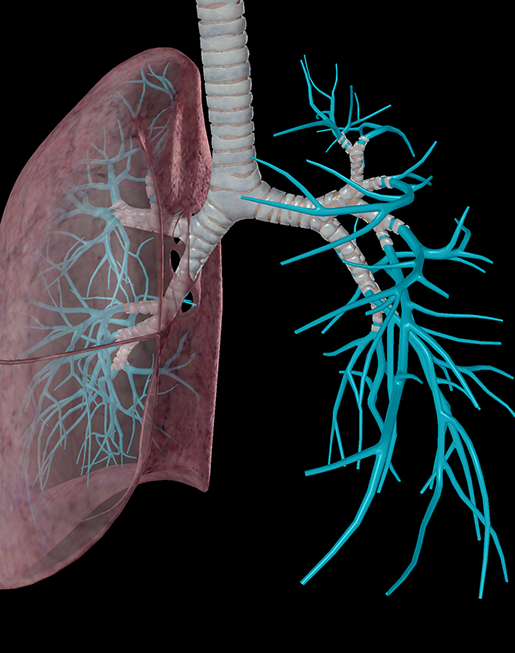
Image from Human Anatomy Atlas.
Though small, the tertiary bronchi are still important! There are about 10 of these relatively narrow airways. They contain very little supportive cartilage and branch into even narrower bronchi—about .2mm in diameter—dividing into even more bronchioles serving the alveoli.
Alveoli
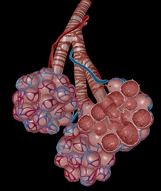
Image from Human Anatomy Atlas.
And here’s where the magic happens (so to speak). Alveoli are tiny, delicate air sacs in which the major and main function of the respiratory system occurs: bringing oxygen into the body and removing carbon dioxide. Tiny pores within the walls of an alveolus allow ventilation with the alveolus next to it. Each lung has about 300 million alveoli. Alveolar sacs are clusters of alveoli, located at the ends of thin airways known as alveolar ducts. The alveoli are surrounded by capillaries.
Pulmonary Capillaries
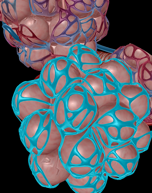 Image from Human Anatomy Atlas.
The pulmonary arteries branch into arterioles, then into networks of capillaries (tiny blood vessels). Gas exchange occurs across the walls of these capillaries, and the oxygen content of the blood within them rises. The pulmonary veins drain the pulmonary capillaries, carrying oxygen-rich blood to the left atrium of the heart for systemic distribution.
… Now for some bonus fun stuff!
Fun Facts About Your Respiratory System
-
You lose a lot of water just by breathing, 17.5 ml or .59 fluid ounces per hour!
-
The average time an adult can hold his or her breath is between 30 and 60 seconds.
-
According to Guinness World Records, the record for a male voluntarily holding his breath is held by Aleix Segura Vendrell from Spain set in 2016. He held his breath for 24 minutes and 3.45 seconds.
-
According to Guinness World Records, the record for a female voluntarily holding her breath is held by Karoline Meyer from Brazil set in 2009. She held her breath for 18 minutes and 32.59 seconds.
-
Pulmonary circulation was first described in the 13th century. Arab physician Ibn al-Nafis described the complicated process in 1243.
And there you have it, the human respiratory system explained in one blog post!
Want to see a 3D tour of the respiratory system? Check out this video!
Video footage from Human Anatomy Atlas.
For even more info about the structures of the respiratory system, take a look at our Respiratory System eBook!
Teaching Respiratory Anatomy with Visible Body Courseware
VB Courseware, Visible Body's teaching and learning platform, comes packed with interactive 3D visual content and sophisticated course management tools that make it easy to teach the respiratory system.
In addition to its library of 3D models, bite-sized learning modules, animations, histology slides, and simulations, Courseware contains a multitude of accessibility tools and features that ensure no student will be left behind. It also has a built-in gradebook that seamlessly integrates with learning management systems like Canvas, Blackboard, Moodle, and more.
To save instructors time and effort, Visible Body has designed premade, customizable curriculum content, including virtual labs and interactive 3D assignments that cover every body system as well as introductory biology topics.

See for yourself! You can get a free instructor trial of Courseware by clicking below.

Be sure to subscribe to the Visible Body Blog for more anatomy awesomeness!
Are you an instructor? We have award-winning 3D products and resources for your anatomy and physiology course! Learn more here.
Additional Sources:


