Anatomy and Physiology: Homologues of Reproductive Anatomy
Posted on 2/21/13 by Courtney Smith
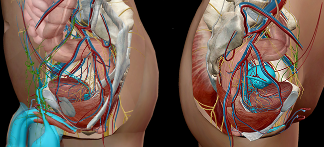 Image from Visible Body Suite.
The battle of the sexes. Men vs. women. Anything you can do, I can do better. For all that battling, it’s amazing how similar we can be. The bits and bobs that everyone seems to think separate us? Really quite similar, actually.
"But," I can hear some of you saying, "there are some obvious differences."
I know what you're thinking… But even if the structures of our reproductive systems look quite different, what if I told you many of their functions are amazingly similar?
And here’s the mind-blowing part: there's a stage in utero in which male and female anatomy is indistinguishable.
Let’s take a look at some similarities between the male and female reproductive systems.
Development of Internal Genitalia (or: Seriously, Our Anatomy Was Exactly the Same At One Point)
There is a time in utero when a developing embryo doesn’t have a noticeable sex. Around 6 weeks, the internal and external genitalia develop, but it’s an in-between state of undifferentiated gender. Think of it like a caterpillar in a chrysalis, undergoing a big change.
An embryo in the early stages (around weeks 5–6) has reproductive structures, ducts, and gonads that can develop into a female or male system. Once the genes determining sex are activated, the appropriate structures will remain while the others degenerate. In the case of a female embryo, it is the paramesonephric (Mullerian) ducts; for the male, it is the mesonephric ducts that develop. The gonads will develop into ovaries or testes.
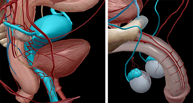
Image from Visible Body Suite.
Development of External Genitalia (or: One of These Things Is Exactly Like the Others)
At the same time of undifferentiated structures in the internal system, the external genitalia is also undifferentiated. No matter our sex, we all start out with the same external anatomy:
|
Genital tubercle |
A protrusion that will develop into the glans penis or the clitoris—both are highly innervated |
|
Urogenital fold |
A structure that becomes the spongy urethra in males or the labia minora in females |
|
Labioscrotal area |
Evolves into the scrotum in males or the labia majora in females |
|
Urogenital membrane |
Ventral part of the cloacal membrane, separating the gut from the external environment |
The images below show the similarities in innervation between the glans penis and the clitoral glans:
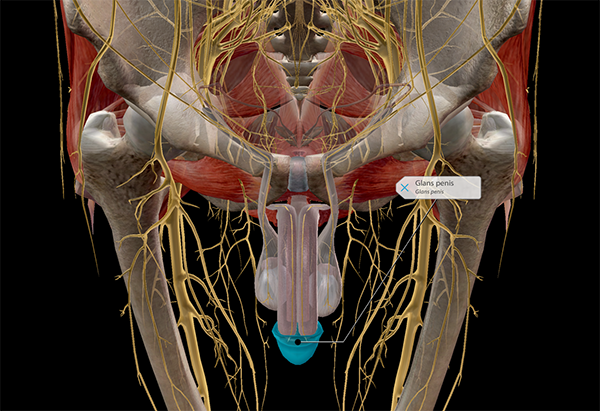
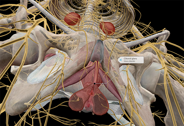
Innervation of the glans penis (m) and clitoral glans (f). Image from Visible Body Suite.
In the next couple of weeks in utero, the undifferentiated anatomy will slowly develop characteristics to match the embryo’s sex.
The Ovaries and Testes (or: I Gots Gonads, You Gots Gonads, Everybody Gots Gonads)
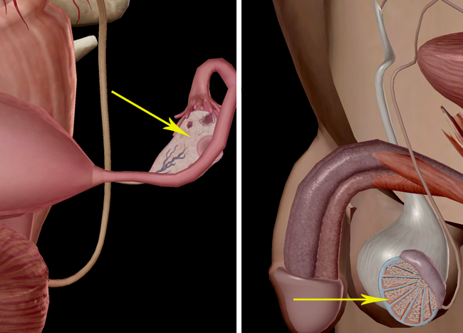
Images from Visible Body Suite.
Ovaries and testes, as I stated before, develop from the same primitive gonads. Even once someone is sexually mature, the testes and ovaries retain almost the exact same egglike shape and function (gamete production). Testes produce about 1,500 sperm/second (see left image). Ovaries contain about two million egg cells (see right image).
The Glans Penis and the Clitoris (or: Stop Giggling)
As any middle schooler knows, the mere mention of these words is enough to give one the case of the giggles. I’ll wait until you’re finished.
Are you done? Awesome.
As I mentioned, in utero we all have a glans area to start with, from which the glans penis or the clitoris eventually develop. Although the penis and clitoris (stop giggling) are different in shape and most functions, what they have in common is nerves. As in, lots of them. There are higher concentrations of nerve endings in the clitoris (8,000+) and the head of the penis (4,000+) than anywhere else in the female and male bodies.
Cowper’s (Bulbourethral) Glands and Bartholin’s Glands (or: I Don’t Have a Cute Subtitle for This)
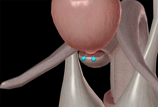
Image from Visible Body Suite.
The human reproductive system has many glands (the male’s more than the female’s), but the bulbourethral glands in males are homologous to the Bartholin’s glands in females. They are both considered accessory glands.
The bulbourethral glands of the male are two pea-sized glands located behind and lateral to the urethra. The gland’s lobules produce alkaline mucus and a single duct carries this mucus into the urethra. The mucus counteracts trace amounts of acid left over from urine, which can interfere with the motility of sperm.
On the other hand, the female’s Bartholin’s glands are two pea-sized, racemose (clustered) glands situated under the skin on either side of the lower part of the vaginal orifice. Like the bulbourethral glands of the male, the Bartholin’s glands secrete a mucus, mostly for lubrication.
There are more homologues than I’ve listed, but those are the big five. So the next time someone tries to lord their gender over you, helpfully remind them that, despite looks, you’re pretty much the same.
Do you want to keep the learning going? Study the structures of the male and female reproductive system with our premade Flashcard decks in Visible Body Suite! Here's how to open them in Atlas:
1. Copy one of these links:
Male reproductive system: https://apps.visiblebody.com/share/?p=vbhaa&t=4_55606_637635904980480000_1048383
Female reproductive system: https://apps.visiblebody.com/share/?p=vbhaa&t=4_55692_637636798136890000_566932
2. Use the Share Link button in VB Suite.
3. Paste the link to view the deck.
4. Save the deck to your User Account.
For more premade Flashcard decks on popular topics, check out our whole library here.
Teaching the reproductive system with Visible Body Courseware
VB Courseware, Visible Body's teaching and learning platform, comes packed with interactive 3D visual content and sophisticated course management tools that make it easy to teach male and female reproductive anatomy and physiology.
In addition to its library of 3D models, bite-sized learning modules, animations, histology slides, and simulations, Courseware contains a multitude of accessibility tools and features that ensure no student will be left behind. It also has a built-in gradebook that seamlessly integrates with learning management systems like Canvas, Blackboard, Moodle, and more.
To save instructors time and effort, Visible Body has designed premade, customizable curriculum content, including virtual labs and interactive 3D assignments that cover every body system as well as introductory biology topics.
 Uterus and endometriosis models in an assignment in Courseware.
See for yourself! You can get a free instructor trial of Courseware here.
Be sure to subscribe to the Visible Body Blog for more anatomy awesomeness!
Are you a professor (or know someone who is)? We have awesome visuals and resources for your anatomy and physiology course! Learn more here.
Related Posts:
- Anatomy and Physiology: Internal Female Reproductive Anatomy
- Anatomy and Physiology: Internal Male Reproductive Anatomy
- Anatomy and Physiology: A look at ovulation, fertilization, and implantation
Additional Sources:
- Visible Body Suite
- Merriam-Webster Dictionary (Bartholin’s gland)
- Tortora, G. (ed.), and Derrickson, B. (2009). Principles of Anatomy and Physiology (12 ed., pp. 1120–1122). Hoboken, NJ: John Wiley & Sons, Inc.


